Whether you’ve received a referral for a PET scan or are exploring your options on your own, we’re here to help you understand what comes next. This page covers everything you need to know about a PET scan—from how to prepare beforehand, to what to expect during the exam, and how to proceed afterward.
What is a PET/MRI scan?
A PET scan shows how your organs and tissues are working, not just what they look like. A PET scan uses a small and safe amount of radioactive substance (called a radiotracer). The radiotracer travels through your body and collects in areas with higher activity—like fast-growing cells or areas of inflammation—highlighting areas of concern. This can help detect disease even before physical changes appear.
PET scans may be performed in combination with MRI scans (PET/MRI) to provide more detailed information about a disease and help guide treatment.
Before Your Appointment
Scheduling Your Exam
Our Patient Access Coordinators are available to assist with scheduling your appointment.
(212) 746-6000
Monday through Friday 8AM to 7PM ET and Saturday and Sunday 9AM to 5PM ET.
Our Locations that Offer PET/MRI
- David H. Koch Center (Upper East Side)
- Weill Greenberg Center (Upper East Side)
Complete Your Exam Forms Easily
Log in to Weill Cornell Connect to complete your safety forms ahead of time using eCheck-In. It’s quick and helps save you time on the day of your appointment.
Don’t have an account? Join today!
Preparing for Your Exam
MRI machines use powerful magnets, so it’s very important to ensure the exam is safe for you. When scheduling and checking in for your appointment, please let our staff know if any of the following apply:
- You have any type of electrical, mechanical, or surgical implant inside your body.
- You have claustrophobia.
- You may be pregnant.
- You have an allergy to contrast dye.
- You have kidney disease (especially if on dialysis).
- You are diabetic.
- You need an interpreter in your language.
Please plan to stay on-site for a minimum of two (2) hours.
FAQ
During Your Appointment
How is a PET/MRI Scan Performed?
- You will lie still on an MRI table that slides into the large, circular opening of the scanning machine.
- As the scan begins, you may hear clicking and banging noises while the images are being captured. Hearing protection will be supplied during all MRIs.
- At certain points during the scan, you may be asked to briefly hold your breath to help capture clear images. During this time, both PET and MRI images will be obtained. The scan will take about 60 minutes.
- Your technologist will be able to see and communicate with you throughout the entire process to ensure you're comfortable.
After the Scan is Completed
The technologist will escort you to your locker to collect your belongings. Our staff will assist you with check-out.
After Your Appointment
Aftercare
- You may be advised to avoid prolonged close contact with pregnant women, babies, or young children for 12 hours after your scan until the radiotracer is fully out of your system.
- You will be instructed to drink 10 glasses of fluids within 24 hours to help flush the radiotracer out of your system.
- You may resume all regular activity unless otherwise directed by your physician.
- While uncommon, if you experience any symptoms of an allergic reaction, please contact us at 646-962-7057.
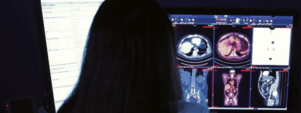
Imaging Review
Once the technologist has taken your images, a radiologist will review the images and write a detailed report. Our radiologists specialize in imaging for specific areas of the body, ensuring you receive the highest-quality, expert interpretation every time.
Our specialties include:
- Abdominal
- Breast
- Cardiothoracic (Heart and Chest)
- Molecular
- Musculoskeletal
- Neuroradiology (Brain, Head, Neck and Spine)
- Pediatrics
Receiving Your Results
Your report will be automatically shared with you and your referring provider within 24-48 hours. You can view your images and reports through Connect. Use this guide to get started.
Sharing Your Results
All imaging exams performed at Weill Cornell Imaging will be available to you in Connect. If you'd like to keep other members of your care team informed, you can easily share your results with any additional providers.
For other medical records requests, visit our Medical Records page.
Understanding Your Results
Please contact your referring provider to discuss your results. They will help explain your imaging report and answer any questions you may have.
If you or your provider have additional questions, our Reading Room Coordinators (RRC) will help connect you with a radiologist.
For more information or to contact an RRC, visit this link.